University of Virginia School of Medicine researchers and their collaborators have solved a decades-old mystery about how E. coli and other bacteria are able to move. Bacteria push themselves forward by coiling long, threadlike appendages into corkscrew shapes that act as makeshift propellers. But how exactly they do this has baffled scientists, because the "propellers" are made of a single protein.
An international team led by UVA's Edward H. Egelman, PhD, a leader in the field of high-tech cryo-electron microscopy (cryo-EM), has cracked the case. The researchers used cryo-EM and advanced computer modeling to reveal what no traditional light microscope could see: the strange structure of these propellers at the level of individual atoms.
"While models have existed for 50 years for how these filaments might form such regular coiled shapes, we have now determined the structure of these filaments in atomic detail," said Egelman, of UVA's Department of Biochemistry and Molecular Genetics.
"We can show that these models were wrong, and our new understanding will help pave the way for technologies that could be based upon such miniature propellers."
Blueprints for Bacteria's 'Supercoils'
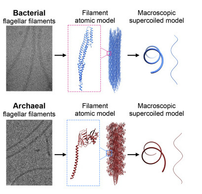
Different bacteria have one or many appendages known as a flagellum, or, in the plural, flagella. A flagellum is made of thousands of subunits, but all these subunits are exactly the same. You might think that such a tail would be straight, or at best a bit flexible, but that would leave the bacteria unable to move. That's because such shapes can't generate thrust. It takes a rotating, corkscrew-like propeller to push a bacterium forward. Scientists call the formation of this shape "supercoiling," and now, after more than 50 years, they understand how bacteria do it.
Using cryo-EM, Egelman and his team found that the protein that makes up the flagellum can exist in 11 different states. It is the precise mixture of these states that causes the corkscrew shape to form. It has been known that the propeller in bacteria is quite different than similar propellers used by hearty one-celled organisms called archaea. Archaea are found in some of the most extreme environments on Earth, such as in nearly boiling pools of acid, the very bottom of the ocean and in petroleum deposits deep in the ground.
Egelman and colleagues used cryo-EM to examine the flagella of one form of archaea, Saccharolobus islandicus, and found that the protein forming its flagellum exists in 10 different states. While the details were quite different than what the researchers saw in bacteria, the result was the same, with the filaments forming regular corkscrews. They conclude that this is an example of "convergent evolution" -- when nature arrives at similar solutions via very different means. This shows that even though bacteria and archaea's propellers are similar in form and function, the organisms evolved those traits independently.
"As with birds, bats and bees, which have all independently evolved wings for flying, the evolution of bacteria and archaea has converged on a similar solution for swimming in both," said Egelman, whose prior imaging work saw him inducted into the National Academy of Sciences, one of the highest honors a scientist can receive. "Since these biological structures emerged on Earth billions of years ago, the 50 years that it has taken to understand them may not seem that long."
Unprecedented details of enamel structure may point to new ways to prevent or halt cavities.
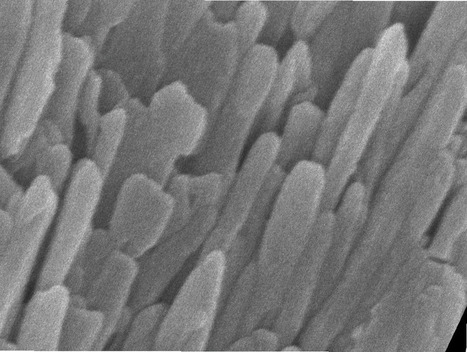
Scientists used a combination of advanced microscopy and chemical detection techniques to uncover the structural makeup of human tooth enamel at unprecedented atomic resolution, revealing lattice patterns and unexpected irregularities. The findings could lead to a better understanding of how tooth decay develops and might be prevented. The research was supported in part by the National Institute of Dental and Craniofacial Research (NIDCR) at the National Institutes of Health. The findings appear in Nature.
“This work provides much more detailed information about the atomic makeup of enamel than we previously knew,” said Jason Wan, Ph.D., a program officer at NIDCR. “These findings can broaden our thinking and approach to strengthening teeth against mechanical forces, as well as repairing damage due to erosion and decay.”
Your teeth are remarkably resilient, despite enduring the stress and strain of biting, chewing, and eating for a lifetime. Enamel — the hardest substance in the human body — is largely responsible for this endurance. Its high mineral content gives it strength. Enamel forms the outer covering of teeth and helps prevent tooth decay, or caries.
Tooth decay is one of the most common chronic diseases, affecting up to 90% of children and the vast majority of adults worldwide, according to the World Health Organization. Left untreated, tooth decay can lead to painful abscesses, bone infection, and bone loss.
Tooth decay starts when excess acid in the mouth erodes the enamel covering. Scientists have long sought a more complete picture of enamel’s chemical and mechanical properties at the atomic level to better understand—and potentially prevent or reverse—enamel loss. To survey enamel at the tiniest scales, researchers use microscopy methods such as scanning transmission electron microscopy (STEM), which directs a beam of electrons through a material to map its atomic makeup.
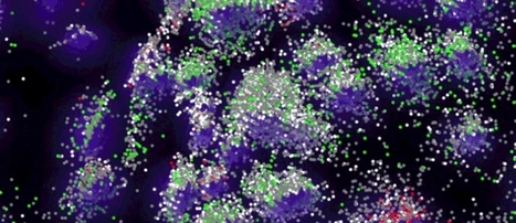
Rather than relying on optics, the DNA microscopy system offers a chemically encoded way to map biomolecules’ relative positions.
Microscopy just got reinvented – again. Traditionally, scientists have used light, x-rays, and electrons to peer inside tissues and cells. Today, scientists can trace thread-like fibers of nerves throughout the brain and even watch living mouse embryos conjure the beating cells of a rudimentary heart. But there’s one thing these microscopes can’t see: what’s happening in cells at the genomic level.
Now, biophysicist Joshua Weinstein and colleagues have invented an unorthodox type of imaging dubbed “DNA microscopy” that can do just that. Instead of relying on light (or any kind of optics at all), the team uses DNA “bar codes” to help pinpoint molecules’ relative positions within a sample.
With DNA microscopy, scientists can build a picture of cells and simultaneously amass enormous amounts of genomic information, Weinstein says. “This gives us another layer of biology that we haven’t been able to see.”
Weinstein, Howard Hughes Medical Institute (HHMI) Investigator Aviv Regev, and molecular biologist Feng Zhang, who was selected as an HHMI investigator in 2018, report the work June 20, 2019, in the journal Cell.
“It’s an entirely new category of microscopy,” Regev says. “It’s not just a new technique, it’s a way of doing things that we haven’t ever considered doing before.”
Until now, insects have been too wriggly to make good subjects for scientists wanting to understand more about insect innards. But an interdisciplinary team of biologists and imaging specialist.
Living insects can now be scanned in unprecedented 3D detail without being harmed. The scanning method developed by scientists at Western University in Canada relies on an insect's ability to survive low-oxygen environments and high-ionizing radiation doses.
Standard methods for looking inside insects involve using dead specimens or killing the bug during the imaging process. "We essentially had snapshots, moments in time, when what we needed were dynamic images of insects' internal development," says biologist Joanna Konopka. "We thought, what would happen if we tried to image them live?"
Konopka therefore teamed up with biophysicist Danny Poinapen to develop a non-invasive technique. Using a steady flow of carbon dioxide, they temporarily immobilized living insects – including Colorado potato beetles and true army worms – for up to seven hours. As the bugs were no longer wiggling around, the team could use X-ray micro-computed tomography (micro-CT) to clearly see the insects' internal workings in 3D. The bugs were then remobilized and the process repeated again days later, with no effect on the insects' longevity, behaviour or ability to reproduce.
The live-scanning method, presented in BMC Zoology, allowed the team to thoroughly examine how insects develop throughout their lifetime. It could therefore be useful in studying parasites or how insects impact human health.
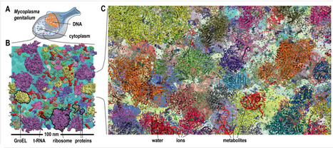
Biological macromolecules function in highly crowded cellular environments. The structure and dynamics of proteins and nucleic acids are well characterized in vitro, but in vivo crowding effects remain unclear. Using molecular dynamics simulations of a comprehensive atomistic model cytoplasm scientists found that protein-protein interactions may destabilize native protein structures, whereas metabolite interactions may induce more compact states due to electrostatic screening. Protein-protein interactions also resulted in significant variations in reduced macromolecular diffusion under crowded conditions, while metabolites exhibited significant two-dimensional surface diffusion and altered protein-ligand binding that may reduce the effective concentration of metabolites and ligands in vivo.
Metabolic enzymes showed weak non-specific association in cellular environments attributed to solvation and entropic effects. These effects are expected to have broad implications for the in vivo functioning of biomolecules.
This work is a first step towards physically realistic in silico whole-cell models that connect molecular with cellular biology.
Two new microscopy techniques are helping scientists see smaller structures in living cells than ever glimpsed before.
Scientists can now view structures just 45 to 84 nanometers wide, Nobel prize-winning physicist Eric Betzig of the Howard Hughes Medical Research Institute’sJanelia research campus in Ashburn, Va., and colleaguesreport in the Aug. 28 Science. The techniques beat the previous resolution of 100 nanometers and shatters the 250 nanometer “diffraction barrier,” imposed by the bending of light.
Using other tricks to improve the super-resolution methods also allowed the researchers to take ultraquick pictures with less cell-damaging light than before. As a result, scientists can watch sub-second interactions within cells, revealing new insights into how cells work.
For years, the lab of Leonard Zon, MD, director of the Stem Cell Research Program at Boston Children’s Hospital, has sought ways to enhance bone marrow transplants for patients with cancer, serious immune deficiencies and blood disorders. Using zebrafish as a drug-screening platform, the lab has found a number of promising compounds, including one called ProHema that is now in clinical trials. But truthfully, until now, Zon and his colleagues have largely been flying blind.
“Stem cell and bone marrow transplants are still very much a black box: cells are introduced into a patient and later on we can measure recovery of their blood system, but what happens in between can’t be seen,” says Owen Tamplin, PhD, in the Zon Lab. “Now we have a system where we can actually watch that middle step.” The animation, based on live imaging of naturally transparent zebrafish, reveals a surprisingly dynamic system in which newborn blood stem cells travel through the blood, exit into a “niche” where they get “cuddled” and nurtured, and then proceed to their final blood-making home. Their journey, also described in the January 15 issue of Cell, offers several clues for helping bone marrow transplants “take.”
“The same process occurs during a bone marrow transplant as occurs in the body naturally,” says Zon. “Our direct visualization gives us a series of steps to target, and in theory we can look for drugs that affect every step of that process.”
Acquista Online La Prescrizione Di Perdita Di Peso Crediamo che i farmaci a volte possano essere molto urgenti da assumere. Se hai urgente bisogno di farmaci, possiamo anche fornirti una consegna espressa,
University of Illinois researchers have developed a new imaging technique that needs no dyes or other chemicals, yet renders high-resolution, three-dimensional, quantitative imagery of cells and their internal structures using conventional microscopes and white light.
Called white-light diffraction tomography (WDT), the imaging technique opens a window into the life of a cell without disturbing it and could allow cellular biologists unprecedented insight into cellular processes, drug effects and stem cell differentiation.
The team, led by electrical and computer engineering and bioengineering professor Gabriel Popescu, published their results in the journal Nature Photonics. “One main focus of imaging cells is trying to understand how they function, or how they respond to treatments, for example, during cancer therapies,” Popescu said. “If you need to add dyes or contrast agents to study them, this preparation affects the cells’ function itself. It interferes with your study. With our technique, we can see processes as they happen and we don’t obstruct their normal behavior.”
Because it uses white light, WDT can observe cells in their natural state without exposing them to chemicals, ultraviolet radiation, or mechanical forces – the three main methods used in other microscopy techniques. White light also contains a broad spectrum of wavelengths, thus bypassing the interference issues inherent in laser light – speckles, for example.
The 3-D images are a composite of many cross-sectional images, much like an MRI or CT image. The microscope shifts its focus through the depth of the cell, capturing images of various focus planes. Then the computer uses the theoretical model and compiles the images into a coherent three-dimensional rendering.
“With this imaging we can tell at what scale things within the cell are transported randomly and at what scale processes are actually organized and deterministic,” Popescu said. “At first glance, the dynamics looks pretty messy, but then you look at it – we stare at movies for hours and hours – and you realize it all makes sense. Everything is organized perfectly at certain scales. That’s what makes a cell alive. Randomness is just nature’s way to try new things.”
Next, the researchers hope to pursue cross-disciplinary collaborations to explore applications of WDT in biology as well as expansions of the imaging optics demonstrated in WDT. For example, they are using WDT to watch stem cells as they differentiate in hopes of better understanding how they turn into different cell types. Since stem cells are so sensitive, only a chemical-free, non-invasive, white-light technique such as WDT could be used to study them without adverse effects.
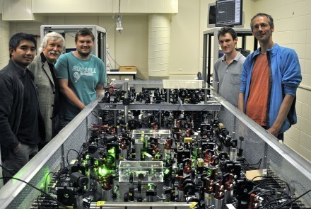
The team, a collaboration between The University of Queensland and the Australian National University, believe their microscope could lead to a better understanding of the basic components of life and eventually allow quantum mechanics to be probed at a macroscopic level. Their world-first discovery has been published in Nature Photonics.
Team leader Associate Professor Warwick Bowen, of UQ’s ARC Centre of Excellence for Engineered Quantum Systems, said the study relied on quantum interactions between the photons of light to achieve measurement precision that surpassed conventional measurement. “This ‘quantum microscope’ is a pioneering step towards applications of quantum physics in technology,” Associate Professor Bowen said.
“In fundamental physics, it could be immediately applied towards observing phenomena in the microscopic motion of small particles that have yet to be observed and were predicted many decades ago.” In the study, the researchers used their quantum microscope to measure the cytoplasm of a live beer-brewing yeast cell and found they could achieve their measurements 64 per cent faster than with a conventional microscope.
Lead author and UQ PhD student Mr Michael Taylor said the results demonstrated for the first time that quantum light could provide a practical advantage in real-world measurements. “The measurements performed could aid in understanding the life-cycle of a cell, as its cytoplasm plays a crucial role in transferring nutrients into and around the cell,” he said.
Among other things, the ‘quantum microscope’ could reveal the finer details within a cell – more than a regular microscope. Biological imaging is a particularly important application for quantum light as these fine details are typically only visible when a lot of light is used.
“Unfortunately, biological samples are grilled when the power is increased too far,” said Mr Taylor. “The ‘quantum microscope’, on the other hand, provides a way to improve measurement sensitivity without increasing the risk of optical damage to the sample.”
By implanting a tiny microscope in the brain of a mouse Stanford researchers have been able to monitor its brain activity.
The study links the observed neuron activity with long-term information storage and could be used to develop treatments and therapies for neurodegenerative conditions in humans.
The technique involved genetically engineering the mice to contain a green fluorescent protein. The protein was created to react to the presence of calcium ions so, when the neuron fired and the cell naturally flooded with those ions, the cells fluoresced green.
A little microscope positioned just above the hippocampus in the mouse's brain could then capture the activity and send it to a computer screen for near real-time monitoring as the mouse runs around a little arena.
"We can literally figure out where the mouse is in the arena by looking at these lights," said biologist Mark Schnitzer, senior author on the paper which has been published in the journal Nature Neuroscience.
"The hippocampus is very sensitive to where the animal is in its environment, and different cells respond to different parts of the arena. Imagine walking around your office. Some of the neurons in your hippocampus light up when you're near your desk, and others fire when you're near your chair. This is how your brain makes a representative map of a space."
These patterns of firing in the mouse brain were found to stay consistent even after weeks had passed between tests. This consistency is what makes it possible to use the technique as a tool with which to study progressive brain diseases and evaluate the effectiveness of some types of treatment and therapy.
The detailed changes in the structure of a virus as it infects an E. coli bacterium have been observed for the first time.
To infect a cell, a virus must be able to first find a suitable cell and then eject its genetic material into its host. This robot-like process has been observed in a virus called T7 and visualized by Ian Molineux, professor of biology in the College of Natural Sciences at The University of Texas at Austin, and colleagues at The University of Texas Health Science Center at Houston (UT Health) Medical School.
When searching for its prey, the virus briefly extends — like feelers — one or two of six ultra-thin fibers it normally keeps folded at the base of its head.
Once a suitable host has been located, the virus behaves a bit like a planetary rover, extending these fibers to walk randomly across the surface of the cell and find an optimal site for infection — the first experimental evidence for this.
At the preferred infection site, the virus goes through a major change in structure in which it ejects some of its proteins through the bacterium’s cell membrane, creating a path for the virus’s genetic material to enter the host.
After the viral DNA has been ejected, the protein path collapses and the infected cell membrane reseals.
“Although many of these details are specific to T7, the overall process completely changes our understanding of how a virus infects a cell,”.said Molineux.
This is also the first time that scientists have made actual images showing how the virus’s tail extends into the host — the very action that allows it to infect a cell with its DNA.
For modern biologists, the ability to capture high-quality, three-dimensional (3D) images of living tissues or organisms over time is necessary to answer problems in areas ranging from genomics to neurobiology and developmental biology. The better the image, the more detailed the information that can be drawn from it. Looking to improve upon current methods of imaging, researchers from the California Institute of Technology (Caltech) have developed a novel approach that could redefine optical imaging of live biological samples by simultaneously achieving high resolution, high penetration depth (for seeing deep inside 3D samples), and high imaging speed.
By blending optical and atomic force microscope technologies, Iowa State University and Ames Laboratory researchers have found a way to complete 3-D measurements of single biological molecules with unprecedented accuracy and precision. Existing technologies allow researchers to measure single molecules on the x and y axes of a 2-D plane. The new technology allows researchers to make height measurements (the z axis) down to the nanometer without custom optics or special surfaces for the samples.
Here's how the technology works: Researchers attach a commercial atomic force microscope to a single molecule fluorescence microscope. The tip of the atomic force microscope is positioned over a focused laser beam, creating a standing wave pattern. A molecule that has been treated to emit light is placed within the standing wave. As the tip of the atomic force microscope moves up and down, the fluorescence emitted by the molecule fluctuates in a way that corresponds to its distance from the surface. That distance can be compared to a marker on the surface and measured.
If you open a biology textbook and run through the images depicting how DNA is organized in the cell's nucleus, chances are you'll start feeling hungry; the chains of DNA would seem like a bowl of ramen: long strings floating in liquid. However, according to two new studies—one experimental and the other theoretical—that are the outcome of the collaboration between the groups of Prof. Talila Volk of the Molecular Genetics Department and Prof. Sam Safran of the Chemical and Biological Physics Department at the Weizmann Institute of Science, this image should be reconsidered. Clarifying it is essential since DNA's spatial arrangement in the nucleus can affect the expression of genes contained within the DNA molecule, and hence the proteins found in the cell.
This story began when Volk was studying how mechanical forces influence cell nuclei in the muscle and found evidence that muscle contractions had an immediate effect on gene expression patterns. "We couldn't explore this further because existing methods relied on imaging of chemically preserved cells, so they failed to capture what happens in the cell nuclei of an actual working muscle," she says.
To address this issue, Dr. Dana Lorber, a research associate in Volk's group, led the design of a device that makes it possible to study muscle nuclei in live fruit fly larvae. The device holds the tiny, translucent larva within a groove that allows it to contract and relax its muscles but keeps its movement constrained so that it can be scanned by a fluorescence microscope. Using the device, the researchers obtained images of the internal, linearly-organized complexes of DNA and its proteins (known as chromatin), surrounded by the membrane of the muscle nuclei.
Expecting a bowl full of ramen, Lorber and Dr. Daria Amiad-Pavlov, a postdoctoral fellow in Volk's group, were in for a surprise. Rather than filling up the entire volume of the nucleus, the "noodles," or long chromatin molecules, were organized as a relatively thin layer, attached to its inner walls. Similar to the outcome of the interaction between oil and water, what is known as "phase separation," the chromatin separated itself from the bulk of the liquid inside of the nucleus and found its place at its outskirts, while most of the fluid medium remained at the center.
The researchers realized that they were on their way to addressing a fundamental biological question, that is—how is chromatin, and hence DNA, organized in the nucleus in a living organism. "But the findings were so unexpected, we had to make sure no error had crept in and that this organization was universal," Lorber says.
Pathology typically involves cutting tissue samples by hand, placing each sample between two pieces of glass, and studying it under a microscope. While microscopes have improved with time, the method has remained largely unchanged for more than 150 years. A human can typically process about 12 sample slices per hour.
3Scan speeds this process up considerably. Its KESM tool uses an automated diamond knife to cut samples at 1,000 slices per hour while simultaneously scanning an image of each slice. Those scans are layered to create a 3D tissue model with micron-scale resolution.
3Scan’s platform has the potential to illuminate the mechanisms by which biological processes become abnormal, which could improve diagnostics, Daniel says: “There’s only so much tissue one person can see in their lifetime, and if we can build something that looks at pathology across many different demographics, across many different cases and diseases, we can get better insights.
There are also reports that show that pathologists achieve consensus on a case 80% of the time. That’s a pretty high success rate—unless it’s your diagnosis, in which case it’s very scary.” Once 3Scan’s tools do the imaging and initial analytics, the data is sent back to pathologists, who explore and translate the findings. “We like to imagine the pathologist as the conductor of an orchestra of robots that can go out there and image vast fields of biology,” Daniel, one of the inventors says. “The pathologist plays a crucial role in being the informed human perspective, differentiating what is pathologic versus normal within that biology.”
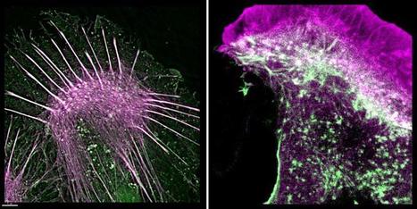
Scientists have developed a new way to see inside individual cells, and study how they move and operate inside the human body. The improved understanding of cell-level activity could give researchers extra insight and tools to tackle cancers and other diseases.
The researchers are using a lab-made protein called an Affimer that binds to the F-actin protein. F-actin is part of the network within cells which gives them their shape and helps them move and divide. By seeing the cells move and change, scientists can begin to develop chemical compounds to target them, over time becoming new drug candidates.
Affimers were first developed at the University of Leeds and are a man-made alternative to animal-derived antibodies. This has the important extra benefit of reducing the numbers of animals used in research work.
This latest type of Affimer technology which recognises the F-actin protein is an important step forward in giving scientists the tools to combat diseases. Details are published in the Scientific Reports journal.
Northwestern University engineers have invented a tool to make a super-high-resolution representation of RNA folding as it is being synthesized. It could potentially lead to future discoveries in basic biology, gene expression, RNA viruses, and disease.
Made up of long chains of nucleotides, RNA is responsible for many tasks in the cellular environment, including making proteins, transporting amino acids, gene expression, and carrying messages between DNA and ribosomes. To accomplish all these tasks, RNA folds into complex structures — “one of the biggest, most essential pieces of biology that we know comparatively nothing about,” said Julius B. Lucks, Ph.D, an associate professor of chemical and biological engineering at Northwestern’s McCormick School of Engineering.
RNA folding is an essential requirement to life, but is difficult to investigate because the process occurs rapidly and is extremely hard to measure. Existing technology to image RNA folding is very-low-resolution and can’t image RNA’s individual components rapidly enough to capture these processes.
Instead, Lucks’s technology combines two existing components: a next-generation sequencing technique, which is typically used for sequencing human genomes, and a chemistry technique to turn RNA structure measurements into big data.
“Instead of treating it like a genome sequencer, we’re treating it like a molecular microscope to get a massive snapshot,” Lucks said. The technique captures the RNA-folding pathway in a massive dataset. Lucks’s group then uses computational tools to mine and organize the data, which reveals points where the RNA folds and what happens after it folds.
From the structural information that they gather, the researchers can reconstruct a movie of the RNA folding process. The team plans to make the data-analysis component open source, so researchers anywhere can download and run the program.
Lucks and his team have already used the technology to view the folding of a ribo-switch, a segment of RNA that acts as a genetic “light switch” to turn protein expression on or off in response to a molecular signal, in this case fluoride.
What looks like a blurry, misshapen flower is actually the innards of one of the world's largest viruses, imaged in three dimensions using powerful X-rays. The same technique could one day create three-dimensional (3D) snapshots of individual molecules, and perhaps even of live bacteria.
The mimivirus (Acanthamoeba polyphaga mimivirus; shown rotating above) carries DNA inside an icosahedral (20-faced) outer shell, and is nearly as large as a typical bacterium. Researchers used the Linac Coherent Light Source (LCLS) at the SLAC National Accelerator Laboratory in Menlo Park, California, to fire powerful X-rays at a single virus particle and build up the 125-nanometre-resolution picture of its internal electron density.
The scans — which are published on 2 March in Physical Review Letters1 — are a proof of principle that extremely powerful X-ray beams could one day take pictures of small objects that cannot be crystallized, says Janos Hajdu, a molecular biophysicist at Uppsala University in Sweden.
Structural biologists routinely fire beams of X-rays at complex molecules and viruses to decode their shapes. But a single molecule does not scatter sufficient X-rays to allow its shape to be reconstructed. In X-ray crystallography, the problem is solved by arranging many copies of the same object into a crystal and looking at repeating patterns in the scattered light. But some molecules are hard to crystallize. And larger, more-complex objects tend to differ from one specimen to the next — for example, genetic material is not arranged in the same way inside all living cells of the same bacterium strain.
The solution could lie in machines known as free-electron lasers, which produce short, densely-packed pulses of X-rays. Each pulse packs in so many high-energy X-ray photons that the machines can — in theory — produce pictures even of single molecules. The LCLS was the first of a handful of such facilities that now exist around the world.
By combining two highly innovative experimental techniques, scientists at the University of Illinois at Urbana-Champaign have for the first time simultaneously observed the structure and the correlated function of specific proteins critical in the repair of DNA, providing definitive answers to some highly debated questions, and opening up new avenues of inquiry and exciting new possibilities for biological engineering.
Scientists who study biological systems at the molecular level have over the years looked to the structure of protein molecules—how the atoms are organized—to shed light on the diverse functions each performs in the cell. The inverse is also true: observing the specific work particular protein molecules perform has provided important clues as to the conformation of the respective molecules. But until recently, our most advanced laboratory experiments could only investigate one at a time—static form or dynamic function—and from the results, deduce the other. This indirect method often doesn’t provide definitive answers.
Now Illinois biological physicists Taekjip Ha and Yann Chemla have combined two cutting-edge laboratory techniques that together directly get at the structure-function relationship in proteins. Ha, who co-directs the Center for the Physics of Living Cells at Illinois, is well recognized for his innovative single molecule fluorescence microscopy and spectroscopy techniques. Chemla is a top expert in optical trapping techniques. Their combined method—simultaneous fluorescence microscopy and optical trapping—yields far more definitive answers to questions relating structure to function than either technique could independently.
Working in collaboration, Ha and Chemla each applied the above techniques in their laboratories, with conclusive results. The findings of these experiments have been published in two separate articles in the April 17 issue of the journal Science.
Sean Carroll's research is directed towards gaining a comprehensive picture of the genetic control of body pattern in fruit flies, butterflies, and other animals. By combining genetic and embryological approaches with molecular techniques for analyzing the structure and expression of key genes that control the fate of groups of cells, we are elucidating the developmental regulatory mechanisms that guide the formation of embryos and body parts. The genes controlling the basic body pattern are fundamental to understanding the origin of the different body structures found in Arthopods and other segmented animals. Using our knowledge of Drosophila, we are studying the developmental and genetic basis of body pattern evolution.
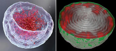
A new method for visualising chromosomes is painting a truer picture of their shape, which is rarely like the X-shaped blob of DNA most of us are familiar with.
Scientists at the BBSRC-funded Babraham Institute, working with the University of Cambridge and the Weizmann Institute, have produced beautiful 3D models that more accurately show their complex shape and the way DNA within them folds up.
The X-shape, often used to describe chromosomes, is only a snapshot of their complexity. See the press release here: http://bit.ly/H68JeH
A dream of scientists has been to visualize details of structures within our cells in real time, a breakthrough that would greatly aid in the study of their function. However, even the best of current microscopes can take minutes to recreate images of the internal machinery of cells at a usable resolution.
Thanks to a technical tour de force, Yale University researchers can now generate accurate images of sub-cellular structures in milliseconds rather than minutes.
This image of microtubules, which act as a cellular scaffolding, was captured in just 33 milliseconds. “We can now see research come to life and tackle complex questions or conditions which require hundreds of images, something we have not been able to do before,” said Joerg Bewersdorf, assistant professor of cell biology and biomedical engineering and senior author of the research, published in the journal Nature Methods.
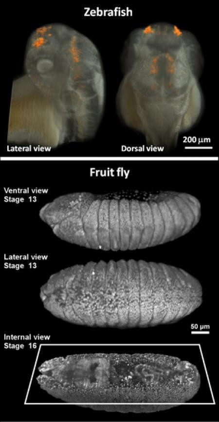
Automated 3‑D analysis of zebrafish larvae, often used as a window on embryonic growth, could aid in the development of new drugs.
Zebrafish larvae — tiny, transparent and fast-growing vertebrates — are widely used to study development and disease. However, visually examining the larvae for variations caused by drugs or genetic mutations is an imprecise, painstaking and time-consuming process.
Engineers at MIT have now built an automated system that can rapidly produce 3D, micron-resolution images of thousands of zebrafish larvae and precisely analyze their physical traits. The system, to be described in the Feb. 12 edition of Nature Communications, offers a comprehensive view of how potential drugs affect vertebrates, says Mehmet Fatih Yanik, senior author of the paper.
“Complex processes involving organs cannot be accurately recapitulated in cell culture today. Existing 3-D tissue models are still far too simple to model live animals,” says Yanik, an MIT associate professor of electrical engineering and computer science and biological engineering. “In whole animals, the biology is far more complicated.”
Lead authors of the paper are MIT graduate student Carlos Pardo-Martin and Amin Allalou, a visiting student at MIT. Other authors are MIT senior research scientist Peter Eimon, MIT intern Jaime Medina, and Carolina Wahlby of the Broad Institute.
I believe this is helpful in many ways. In the science world, we need as many things we can discover. The more we know, the more we can work with. There are so many new opportunities now because we can look into the complications of traits in animals. Also, zebra fish can be used as an aid of developing new drugs and medicines.
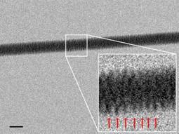
It's the most famous corkscrew in history. Now an electron microscope has captured the famous Watson-Crick double helix in all its glory, by imaging threads of DNA resting on a silicon bed of nails. The technique will let researchers see how proteins, RNA and other biomolecules interact with DNA.
The structure of DNA was originally discovered using X-ray crystallography. This involves X-rays scattering off atoms in crystallised arrays of DNA to form a complex pattern of dots on photographic film. Interpreting the images requires complex mathematics to figure out what crystal structure could give rise to the observed patterns.
The new images are much more obvious, as they are a direct picture of the DNA strands, albeit seen with electrons rather than X-ray photons. The trick used by Enzo di Fabrizio at the University of Genoa, Italy, and his team was to snag DNA threads out of a dilute solution and lay them on a bed of nanoscopic silicon pillars.
The team developed a pattern of pillars that is extremely water-repellent, causing the moisture to evaporate quickly and leave behind strands of DNA stretched out and ready to view. The team also drilled tiny holes in the base of the nanopillar bed, through which they shone beams of electrons to make their high-resolution images. The results reveal the corkscrew thread of the DNA double helix, clearly visible. With this technique, researchers should be able to see how single molecules of DNA interact with other biomolecules.
The growing class of fluorescent proteins useful for detecting events in living cells and animals has almost single-handedly launched and fueled a new era in biology and medicine.
It took over thirty years, and the advent of recombinant DNA as well as vastly improved molecular biological approaches to see the pioneering work of Osamu Shimomura developed into a useful tool for live-cell imaging by Doug Prasher and Martin Chalfie. Just in the past decade, however, we have witnessed a truly remarkable expansion in the fluorescent protein palette, largely driven by the innovative studies from Roger Tsien's laboratory. Most of the fluorescent proteins that are commonly used today have been modified through mutagenesis to optimize their expression in biological systems. Continued efforts using directed evolution approaches will no doubt improve the spectral characteristics, photostability, maturation time, brightness, acid resistance, and utility of the fluorescent protein tags for cellular imaging.
The current thrust of fluorescent protein development strategies is centered on fine-tuning the current palette of blue to yellow variants from jellyfish, while simultaneously developing monomeric fluorescent proteins emitting in the orange to far-red regions of the visible light spectrum. We now have jellyfish proteins that span an 80-nanometer portion visible spectrum from deep blue to yellow-green, providing a wide choice of genetically encoded markers for studies in cell biology. Fluorescent proteins derived from Anthozoa species (corals and anemones), as well as other sources, span the entire visible spectrum and feature a wide range of useful properties. The unique optical highlighter properties of fluorescent proteins can allow the investigator to change the color or the emission state, providing unique opportunities to track the dynamic behavior of proteins in living cells and animals.
Still the "gold standard" in fluorescent protein technology, the enhanced version of GFP features a chromophore based on a para-hydroxybenzylidene substituted imidazolinone. The chromophore of the first reported red fluorescent protein extends conjugation into the polypeptide backbone to generate fluorescence in the longer wavelength regions. The ZsYellow fluorescent protein chromophore features a novel three-ring system and peptide backbone cleavage due to the substitution of lysine for serine as the first amino acid residue in the chromophore tripeptide. The final step in mKO chromophore maturations involves the formation of a novel five-member thiazole ring system when the Cys65 hydroxyl moiety attacks the carbonyl of Phe64 and cyclizes. In a manner similar to mKusabira Orange, mOrange chromophore maturation involves the formation of a novel five-member oxazole (rather than a thiazole) ring system.
To get content containing either thought or leadership enter:
To get content containing both thought and leadership enter:
To get content containing the expression thought leadership enter:
You can enter several keywords and you can refine them whenever you want. Our suggestion engine uses more signals but entering a few keywords here will rapidly give you great content to curate.


 Your new post is loading...