Your new post is loading...

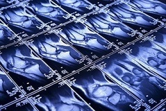

Combining multiple medical images from one patient can provide important information. This is not always easy to do with the naked eye. This is why we need software that can compare different medical images. To do so, so-called image registration methods are used, which basically compute which point in one image corresponds to which point in another image.
Current solutions are often not always suitable for use in a medical setting, which is why AMC and CWI together with companies Elekta and Xomnia will develop a new image registration method.
Suppose you have multiple CT and/or MRI images of a patient, made at different points in time. Medical staff wants to compare these images, for example to see how certain irregularities have developed over time. But these images are often fundamentally different (e.g., patients never lie in a scanner in the exact same manner) and when different imaging methods are used this is even more complicated.
So how can one determine precisely what has changed?
With the software that is currently available this can be very hard, or even impossible, to accomplish in practice.
This project has 2 major challenges. The models and algorithms for large deviations have to be improved. Next to that, the software has to be designed so that it is intuitive to use and helps medical practitioners get the results they want. By combining new deformable image registration models and algorithms with machine learning, the software can be trained on example cases to work even better. The focus of the project will be on supporting better radiotherapy treatment, with validations in the real world (i.e., the clinic), but the method will also be applicable to other (medical) areas.
more at https://www.cwi.nl/news/2017/comparing-medical-images-better